- AdventHealth
Anatomage Table enhances teaching anatomy and increases student learning at AHU
This Physician's View opinion piece is written by Mohtashem Samsam, M.D., Ph.D., professor at AdventHealth University (AHU) and director of research at AHU.
During my medical school years, cadavers were the only opportunity to learn about the human anatomy. We also used anatomy atlases and other books to study and learn the intricoes of what we would eventually dissect. In my experience, one of the best ways of teaching 3D anatomy is through dissection or prosection of the human body, but thanks to technology and innovation in virtual anatomy including the Anatomage Table, there are now more options in studying the human body.
The New Age of Dissection
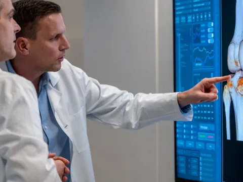
Standing nearly seven feet in length and displaying images in horizontal or vertical positions, the Anatomage Table is a 3D virtual anatomy, physiology and pathology device (think of an enlarged iPad, larger than the height of an average American man) that uses color images, CT scans and other images of donated cadavers.

From an instructor perspective, the table allows students to learn independently, which considerably cuts down learning and teaching time.
Moving layer by layer when virtually dissecting the body, a student can make precise incisions with their fingertips and rotate the image to see cross-sections, just as they would in a CT scan. Unlike a cadaver, which can be limited in viewpoints once a cut is made, this technology allows the student to examine the body in various sections – transverse, sagittal and coronal – as many times as desired.
In addition, digital images used in the table dissections are from real-life patient scans, which means each person is unique. The images also offer each student an amazingly accurate sense of depth, which is important for surgery and procedural interventions and manipulative therapies.
Benefits of 3D Dissection
Anatomy education through human cadavers is essential for students’ learning and practice, especially in surgery, interventional areas, physical medicine, physical therapy and others. However, since installing this technology at AdventHealth University (AHU) about a year and a half ago, my colleagues and I have found that 3D virtual anatomy dissection, including the use of the Anatomage Table, enhances the education experience and help students’ learning.
As another benefit, the structure of cadavers isn’t always in the best shape after a couple of weeks after dissection – the superficial tissues and organs may not be in the right location or orientation – but with the Anatomage Table, you can dissect and repeat as many times as you want. This is ideal for students in their basic science education as they start their training, as well as later in clinical trainings in specific areas.
The Evolution of 3D Tables in Clinical Training
Collaboration of the AdventHealth Nicholson Center, an international educational institute for medicine with AHU, provides a unique dissection opportunity and the Anatomage Table is an opportunity for AHU to continue offering leading-edge technology and equipment to students.
Because this technology provides students a glimpse into pathology and physiology, such as changes in heart function, it may have future uses as a tool for research and case-based simulations in pathology. For example, what is happening to a person’s heart rate while an incision is being made, or how does a brain disorder affect the muscles in one’s legs?
On the Anatomage Tables...

Mohtashem Samsam, M.D., Ph.D., professor at AdventHealth University (AHU) and director of research at AHU


